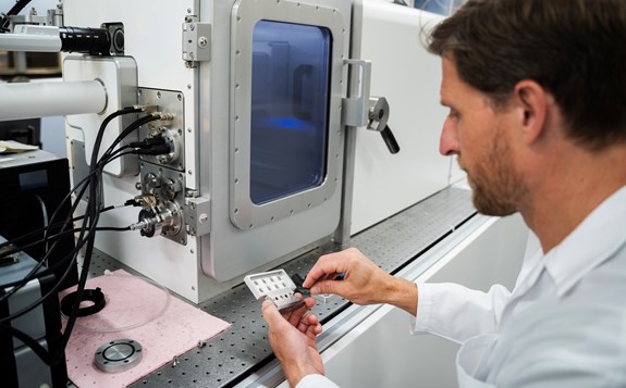
Imaging
More about Nikon Eclipse LV100ND light microscope
- The microscope is equipped with a Linkam Analysa-LTS350 temperature-controlled stage to allow imaging of samples at elevated temperatures, for example to investigate temperature-induced phase transitions. It is further equipped with polarising filters for both reflected and transmitted light, to allow study of birefringent samples. There are two sets of objectives, one for short working distance (10X, 40X, 100X) for imaging of flat samples with cover glass; and one set with long working distance (10X, 50X), for imaging the surface of non-flat samples or when using the temperature-controlled stage. A TransferMan NK2 micromanipulator allows manipulation of samples during imaging, such as mounting of AFM colloidal probes. The microscope is equipped with a CAM-UC90, 9 megapixel, 1” CCD, 4k resolution digital camera and is running cellSens Entry V2.
Specifications
- Upright light microscope
- LED illumination for both transmitted and reflected light
Available objectives:
- CFI Plan Akromat 10X, N.A. 0.25, W.D. 10.5 mm
- CFI Plan Akromat 40X, N.A. 0.65, W.D. 0.56 mm
- CFI Plan Akromat 100X olja, N.A. 1.25, W.D. 0.2 mm
- CFI TU Plan Fluor Epi 10X, N.A. 0.30, W.D. 17.5 mm
- CFI TU Plan Epi ELWD 50X, N.A. 0.60, W.D. 11 mm
Special features:
- Temperature-controlled stage Linkam Analysa-LTS350
- TransferMan NK2 micromanipulator
- Polarising filters for both reflected and transmitted light
- Acquisition Software: cellSens Entry V2

More about Evident FLUOVIEW FV5000 confocal laser scanning microscope
Specifications
Laser lines: 405 nm, 445 nm, 488 nm, 561 nm, 594 nm, 640 nm
Detectors:
- 4 low-noise SilVIR type detectors (2 blue sensitive, 2 red sensitive)
- 1 transmitted light detector
Scanners:
- 8kx8k Galvanometric scanner, for best quality images
- 2kx2k Resonance scanner, for fast imaging
Available objectives:
- UPLXAPO 4X NA 0.16 (dry)
- UPLXAPO10X O NA 0.40 WD 3.1mm (dry)
- UPLXAPO 20X NA 0.80 WD 0.6mm (dry)
- UPLAPO 25x NA 1.0 WD 1.0mm
- UPLXAPO 40x NA0.95 WD0.18 (dry)
- UPLSAPO 60xOHR NA1.50 WD 0.11 (oil, highest numerical aperture)
- PLAPON 60XOSC2 NA 1.40 WD 0.12 (oil, super-corrected, for co-localisation studies)
Acquisition Software: cellSensFV
Special Features:
- Stage-top climate control chamber Okolab UNO+
- LED fluorescence light source for transmitted and reflected light
- High-resolution camera for wide-field fluorescence imaging or non-fluorescent staining
- Software-controlled auto-correction collar for one objective
- Auto-adjusted laser intensity
- Auto sample finder
- Auto-focus finder with hardware-based z-drift compensator
- AI-supported de-noising functions
- Live and post-processing deconvolution functions
- FV-OSR Super resolution module for X-Y imaging down to 120 nm

More about Zeiss EVO LS-10 scanning electron microscope
By using different types of detectors, different types of signals can be collected, revealing complementary information about the sample. The standard secondary electron detector will give the best signal-to-noise and thereby the best image quality. The backscatter detector detects only high-energy backscatter electrons, potentially originating from deeper into the sample, which gives a flatter and less detailed image, but additionally offers information about sample composition, as differences in intensity will indicate differences in atomic weight of the exited elements. By collecting X-ray radiation generated by the sample, the EDX (electro-dispersive X-ray) detector can identify and quantify the relative composition of the elements in selected parts of the sample, either for larger areas (entire part of the sample currently imaged), point and ID (for selected parts/features of the sample) or by mapping.
The Zeiss EVO LS10 is an environmental scanning electron microscope, equipped with a LaB6 filament. The EVO LS10 offers two additional imaging modes, apart from the standard high vacuum mode for fully conducting samples. In variable pressure mode, a low pressure of nitrogen gas (usually 10-40 Pa) is allowed in the chamber. The gas is ionised by the electron beam and the ions help dissipate the surface charge built up in poorly conducting samples, allowing imaging of non-conductive samples without surface coating. In extended pressure mode, water vapour is introduced into the chamber, which, together with a Peltier Coolstage, allows imaging of hydrated samples at controlled temperature and relative humidity.
An Agar automatic sputter coater is also available for gold coating of samples.
Specifications
- LaB6 filament, 1-30 kV
Detectors:
- Everhart-Thornley Secondary electron detector
- Lens mounted 4Q-BSD backscatter detector
- VPSE G3 (Variable pressure secondary electron detector)
- INCA x-act LN3-free EDX detector
Imaging modes:
High vacuum
- Best image quality, for conductive or gold-coated samples
Variable pressure
- Lower image quality, for non-conductive samples where coating is impossible or unwanted
- 10-40 Pa of nitrogen gas in the sample chamber
Extended pressure
- For cooled, hydrated samples such as cells/bacteria, skin, polymers
- Temperature-controlled Peltier stage
- 1-3000 Pa of water vapour in the sample chamber, allows controlled relative humidity
Sample holder and stage:
- 8-position sample carousel for Zeiss 13 mm pin-type sample stubs
- Multi-purpose quick-fit sample holder for larger irregular specimens
- Maximum specimen height 100 mm, maximum diameter 230 mm
- 5-axis stage: X, Y, Z, rotation (0-360) and tilt (0-90)